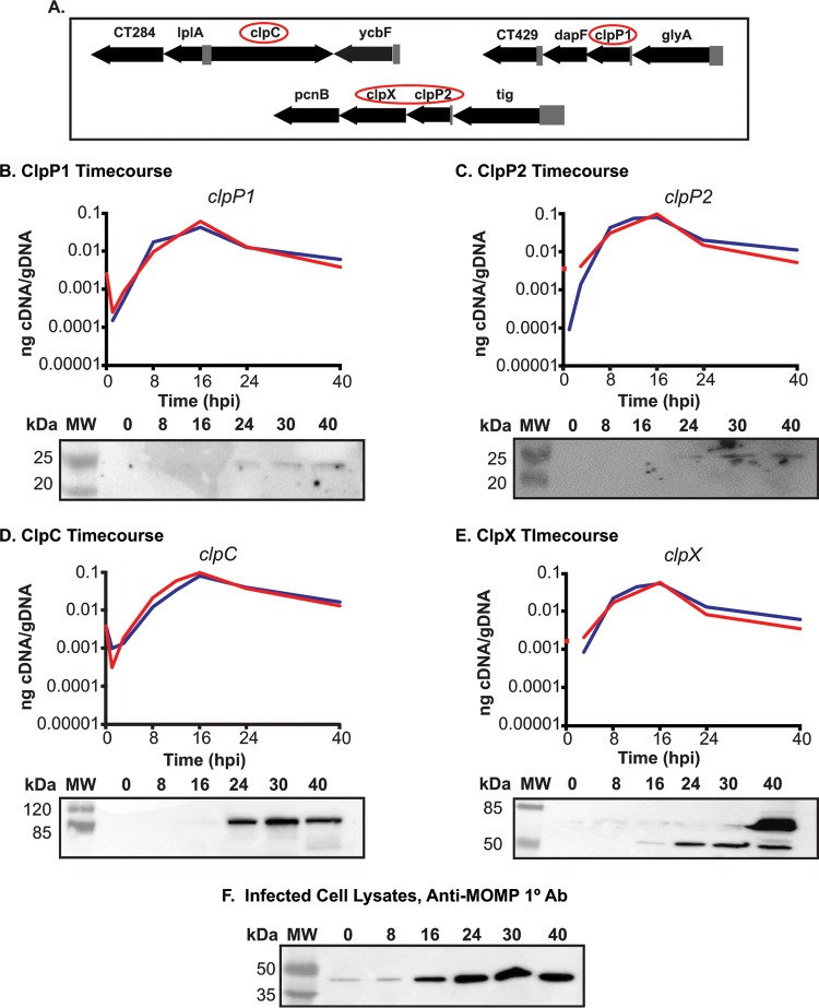
FIG 1.
The clp genes are expressed during RB growth. (A) Gene maps of the clp genes in the Chlamydia trachomatis genome. Gene numbers are in numerical order from left to right and reflect the serovar D numbering scheme from a study by Stephens et al. (16). The clp genes are circled. The clpP2 gene is ct706. (B to E) Temporal expression of the clp genes. Quantitative reverse transcription-PCR (RT-qPCR) analysis of the clp genes from two independent time course experiments of a C. trachomatis serovar L2 infection of HEp2 cells was performed. Total RNA and DNA were collected at the indicated times postinfection and processed as described in Materials and Methods. Equivalent amounts of cDNA were used for each assay and analyzed in triplicate. Results are reported as a ratio of cDNA to genomic DNA (gDNA). Standard deviations for each were typically less than 10% of the sample. Note that some transcripts were not detectable at 1 hpi. Western blotting was performed on whole-cell lysates of total protein from a time course of Chlamydia sp.-infected cells, separated by SDS-PAGE, and transferred to a nitrocellulose membrane for blotting. All four of the genes analyzed appear to be expressed mid-developmental cycle. (F) Major outer membrane protein (MOMP) was blotted as a control for chlamydial development over the course of infection. Ab, antibody.

