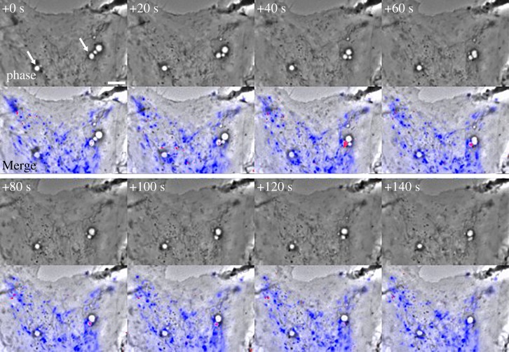
Figure 2.
Solute size-dependent delivery of dye from endolysosomes into macropinosomes by piranhalysis. Time-series of a macrophage stimulated with M-CSF after pre-loading endolysosomes with Lucifer yellow (LY) and Texas Red-labelled dextran (TRDx) (see also electronic supplementary material, Video S2). One image set was collected every 20 s. Top panels: Phase-contrast. Indicated times, in seconds, are relative to the first frame. Arrows at +0 s indicate two macropinosomes which received LY and TRDx during the sequence. Bottom panels: phase-contrast with a blue overlay indicating endolysosomes containing both LY and TRDx fluorescence and red overlay indicating macropinosomes containing increased concentrations of LY relative to TRDx. Preferential labelling of macropinosomes with LY indicated the molecular size-selective transfer of fluid-phase probes between the interacting organelles (i.e. LY entered macropinosomes from endolysosomes earlier than did TRDx). Scale bar: 5 µm. Adapted from a supplementary movie described in Yoshida et al. [6].

