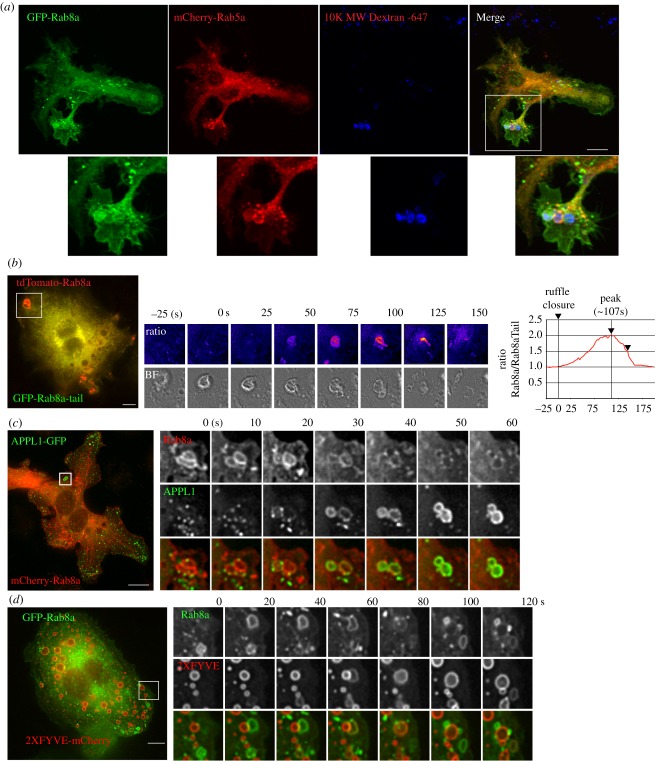
Figure 2.
Rab8a recruitment analysis during macropinosome formation. (a) RAW 264.7 cells transiently expressing GFP-Rab8a and mCherry-Rab5a. Cells were pulsed for 15 min with 647-dextran to label newly formed macropinosomes imaged on a Zeiss AxioImager. (b) Ratiometric time-lapse imaging of full-length tdTomato-Rab8a and GFP-Rab8a-Tail domain. Right shows ratio of two channels compared to formation of the macropinosome, depicted by brightfield image. Time-lapse imaging of cells co-expression of (c) APPL1-GFP and mCherry-Rab8a and (d) GFP-Rab8a and 2xFYVE-mCherry. Cells in (b) and (c) were imaged using the DeltaVision Deconvolution microscope. Scale bar, 10 µm.

