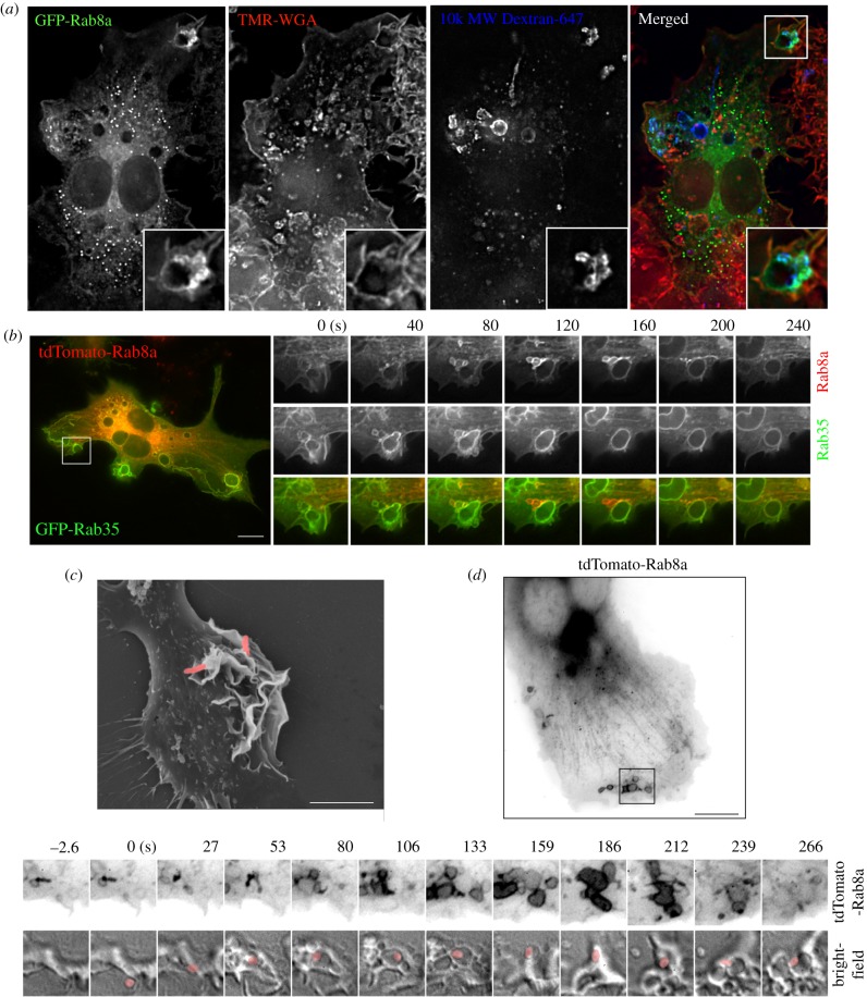
Figure 4.
Rab8a recruitment to phagosome-associated macropinosomes. (a) Medium containing IgG opsonized 3 µm latex beads was added to RAW 264.7 macrophages transiently expressing GFP-Rab8a for 10 min before fixation. Cells were counterstained with TMR-WGA without permeabilisation. (b) RAW 264.7 cells transiently expressing tdTomato-Rab8a and GFP-Rab35 were imaged after addition of IgG opsonized sheep red blood cells. (c) SEM of RAW 264.7 macrophages after infection with Salmonella enterica. Salmonella is pseudo-coloured pink. Scale bar, 5 µm. (d) Live-cell imaging of S. enterica infection of RAW 264.7 macrophages transiently expressing tdTomato-Rab8a. Still image and inset frames are inverted and represented in grey-scale. Salmonella is pseudo-coloured pink. Scale bar, 10 µm.

