Figure 6.
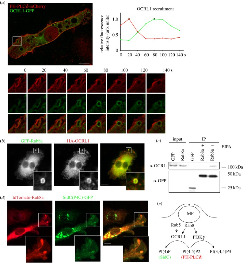
OCRL1 interaction with Rab8a on macropinosomes. (a) RAW 264.7 macrophages transiently expressing PH-PLCδ-mCherry with OCRL1-GFP. Inset shows example of a ruffle transitioning to a macropinosome. OCRL1 recruitment analysis is displayed in arbitrary units and represented relative to each channels' peak intensity. (b) Co-expression of GFP-Rab8a and HA-OCRL1 in RAW 264.7 macrophages. Cells were imaged in (a,b) using the DeltaVision deconvolution microscope. (c) Co-immunoprecipitation analysis of RAW 264.7 macrophages, transiently expressing GFP-Rab8a or GFP-empty vector as a control. Cells were treated with or without 25 µM EIPA to block macropinocytosis and immunoprecipitations were performed using GFP-nanotrap agarose. Immunoblots were probed for anti-GFP and anti-OCRL. (d) Live-cell imaging of RAW 264.7 macrophages transiently expressing tdTomato-Rab8a and SidC(P4C)-GFP. Cells were imaged using Zeiss Axiovert 200 spinning disk microscope. (e) Summary diagram illustrating two possible macropinosome-associated Rab8a effectors that act on PI(4,5)P2.

