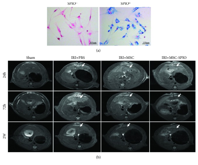
Figure 4.
MSCs accumulated in injured liver lobes in the model of hepatic IRI. (a) SPIO-labeled MSCs were positive for Prussian blue staining. Blue particles were observed in the cytoplasm of SPIO+ cells. (b) Male SD rats were randomized into sham, IRI + PBS, IRI + MSC, and IRI + MSC-SPIO groups. Representative images of MRI scanning on the T2 sequence of I/R lobes in each group were shown after 24 h, 72 h, and 2 w of reperfusion. The liver lobes experienced IRI were pointed out by white arrows. Original magnification, ×200.

