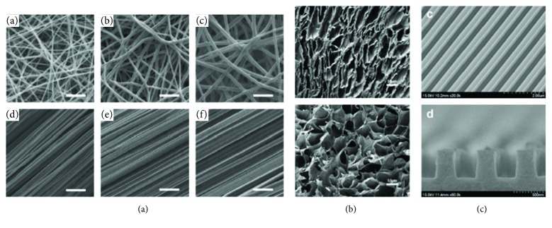
Figure 3.
SEM images of different topographies used to influence PSC fate. (a) Electrospun meshes with differently sized fiber diameters, in random or aligned fiber orientation, investigated with respect to germ layer commitment [73]. (b) Rod (top) or sphere (bottom) pore-shaped scaffold used for bone differentiation. Study found spherical pores supported osteogenic fate better than rod shapes [1]. (c) Nanosize surface grooves instructed PSCS into neuronal lineage without additional inducing agents [78].

