Fig. 3.
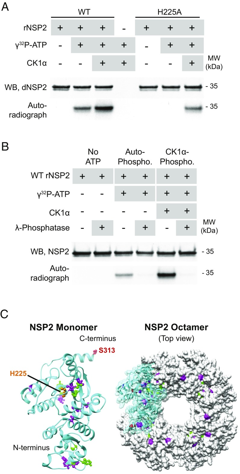
NSP2 autophosphorylates and is phosphorylated by CK1α. NSP2 was separated in reducing gels and detected by WB with anti-dNSP2 monoclonal antibody. Blots were exposed to autoradiography film overnight to detect the transfer of radiolabeled phosphate to NSP2. (A) Phosphorylation of WT (Left) and mutant H225A (Right) rNSP2 in the presence of γ[32P]ATP and CK1α. (B) Effect of lambda-phosphatase treatment on rNSP2, autophosphorylated rNSP2, and CK1α-phosphorylated rNSP2. (C) Crystal structure of WT NSP2 monomer (cyan; PDB ID code 1L9V) and octamer, highlighting autophosphorylated residues (magenta), CK1α-phosphorylated S313 (red), additional autophosphorylated residues detected after CK1α-phosphorylation (green), and H225 (orange).

