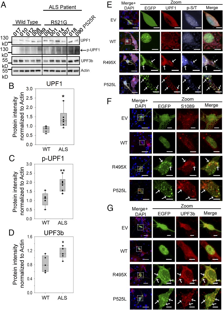
Fig. 3.
Up-regulation of pro-NMD factors in cells of patients with familial ALS and in primary neurons expressing mutant FUS. (A–D) Levels of pro-NMD factors in patients with ALS carrying the R521G or P525R mutation and in control subjects with WT FUS. Western blots of UPF1, p-UPF1, UPF3b, and actin control were performed (A), and quantification of UPF1 (B), p-UPF1 (C), and UPF3b (D) was normalized against actin and obtained from three replicates. Error bars represent the SD between individuals. Quantifications were compared with healthy controls using a Student’s t test. *P ≤ 0.05; **P ≤ 0.005. (E–G) Immunofluorescent staining of UPF1, p-UPF1, and UPF3b in mouse primary neurons transfected with EV, EGFP-tagged WT, or mutant FUS at day 4 of in vitro culture. (E) Immunofluorescent staining of UPF1 and p-UPF1 using an anti–p-S/T ATM/AMR substrate antibody. (F) Immunofluorescent staining of p-UPF1 using an antibody against phosphor-S1089 in UPF1. (G) Immunofluorescent staining of UPF3b. Arrows indicate inclusions where proteins of interest are colocalized. (Scale bars: regular views, 20 μm; zoomed-in views, 5 μm.)

