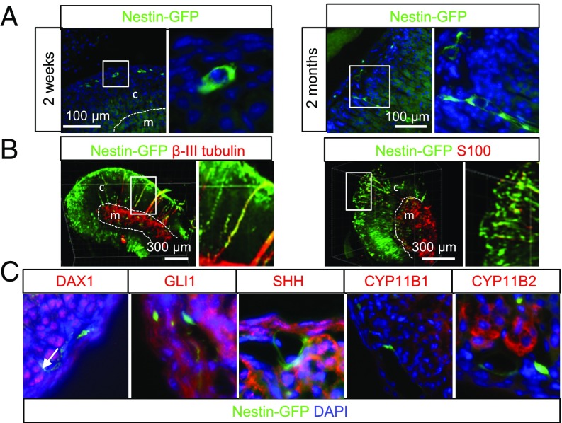
Fig. 1.
Nestin-GFP–positive cells in the adrenal cortex. (A) Localization of Nestin+ cells in young (P14) and adult (2 mo old) mice. (B) Three-dimensional imaging of cleared adrenal sections costained with antibodies against β-III tubulin or S100. (C) Costaining with a panel of known progenitor/stem cell markers and steroidogenic enzymes. Double-positive cells are marked with arrows. Dashed lines mark the border between the cortex (c) and medulla (m).

