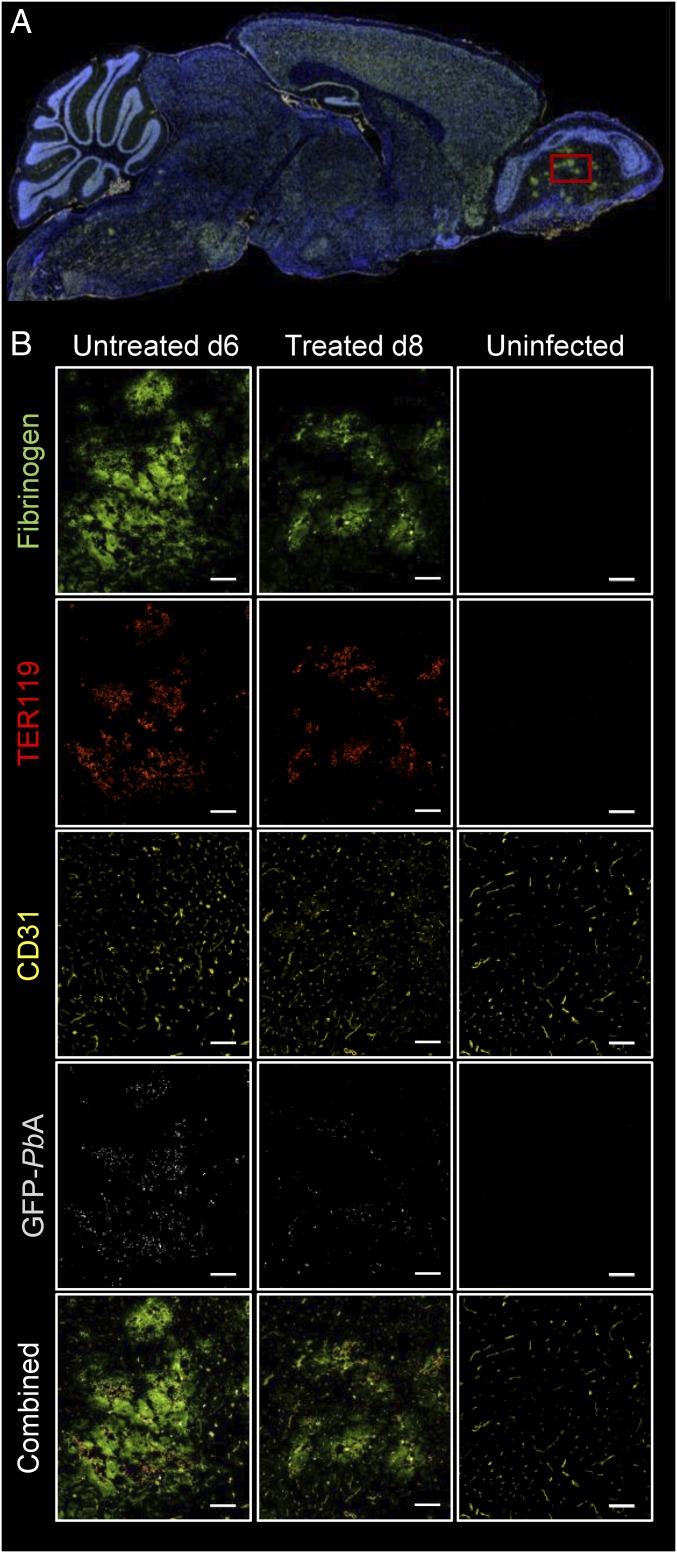
Fig. 7.
Immunofluorescent staining of sagittal sections of mice infected with PbA (untreated and treated) and control uninfected mice. (A) A representative full sagittal section of the stained mouse brain. (B) Magnified images obtained in the olfactory bulbs (red square in A) of an infected untreated mouse that was euthanized 6 d p.i. (Left column), a treated mouse euthanized 8 day p.i. (Middle column), and an uninfected mouse (Right column). Positive staining for fibrinogen (bright green), red blood cells (TER119, red), endothelial cells (CD31, yellow), and GFP-labeled parasites (light gray) show evidence of extravascular protein and blood extravasation in both infected mice compared with the control animal. Regions of protein accumulation colocalize with extravasated, parasite-infected RBCs (33.3% magnification). (Scale bar: 100 µm.)

