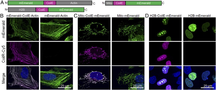
Fig. 2.
Selective fluorescent labeling of cellular actin, mitochondria, and the nucleus using VIPER. (A) Representation of the CoilE-tagged proteins used to label the cytoskeleton (mEmerald-CoilE-Actin), mitochondria (Mito-CoilE-mEmerald), and the nucleus (H2B-CoilE-mEmerald). Transfected U-2 OS cells were labeled postfixation by treatment with CoilR-Cy5 (100 nM) and then imaged by confocal FM to observe the cytoskeleton (B), mitochondria (C), or the nucleus (D). CoilR-Cy5 labeling was specific for CoilE-tagged proteins, and the Cy5 (magenta) and mEmerald (green) signal colocalized. Green-magenta overlap appears white in the merged images and the nuclear stain (Hoechst 33342) is false-colored blue.

