Fig. 6.
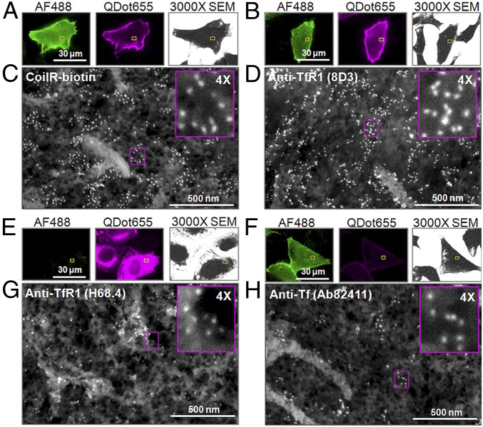
Target labeling and CLEM imaging by VIPER or immunolabeling. Cells expressing TfR1-CoilE were identified by binding to Tf-AF488. For VIPER labeling, fixed cells were treated with CoilR-biotin and streptavidin-Qdot655 (A). For immunolabeling, cells were treated with a primary antibody against Tf (Ab82411; F) or TfR1 [8D3 (B) or H68.4 (E)]. Primary antibodies were detected using secondary antibodies conjugated to Qdot655. Fluorescence micrographs of cells labeled with VIPER (A), 8D3 (B), H68.4 (E), and Ab82411 (F) were acquired and mapped for high-resolution SEM. After processing, we selected regions (yellow boxes) for SEM imaging at 100,000× magnification. The high-resolution view shows Qdot labeling of the cell surface (C, D, G, and H). Magenta Insets present a 4× magnification of the Qdots. For H68.4, detergent treatment caused membrane extraction, as observed by 3000× SEM, and the Tf-AF488 signal was reduced.

