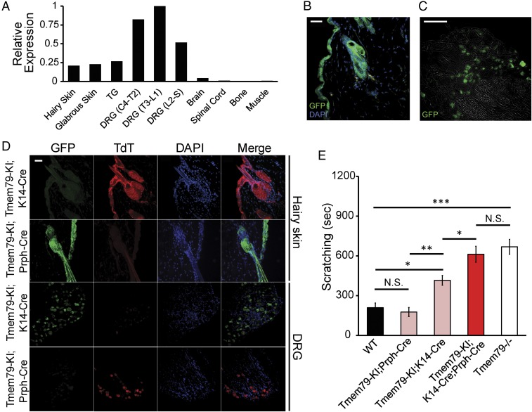
Fig. 1.
Loss of Tmem79 in sensory neurons and keratinocytes produces itch. (A) Real-time qPCR analysis of Tmem79 expression relative to Rpl19 in tissues dissected from wild-type mice. The graph presents ΔΔCt values normalized to the tissue with greatest relative expression of Tmem79. ΔΔCt values were calculated from real-time PCR reactions run in triplicate from combined tissues from three wild-type mice. Tissues examined were hairy skin, glabrous (hairless) skin, TGs, DRG fourth cervical to second thoracic (DRG C4–T2), DRG third thoracic to first lumbar (DRG T3–L1), DRG second lumbar to sacral (DRG L2–S), brain, spinal cord, bone, and muscle. (B and C) Representative micrographs of tissue sections from Tmem79-KI reporter mice stained for immunoreactivity to GFP (green). (B) Hairy skin section (20 µm) with the superficial epidermis oriented to the left. Nuclei were counterstained using DAPI (blue). (Scale bar: 15 µm.) (C) DRG section (12 µm). The image is superimposed with brightfield micrograph. (Scale bar: 100 µm.) (D) Representative micrographs of hairy skin or DRG sections from Tmem79-KI;K14-Cre or Tmem79-KI;Prph-Cre mice stained for immunoreactivity to GFP (green) or TdT (red). Superficial epidermis is oriented to the left (1st Row, Tmem79-KI;K14-Cre) or the right (2nd Row, Tmem79-KI;Prph-Cre). Nuclei were counterstained with DAPI (blue). Merged images are shown in the far right column. (Scale bar: 15 µm.) (E) Quantified scratching behavior of adult wild-type, Tmem79-KI;Prph-Cre, Tmem79-KI;K14-Cre, Tmem79-KI;K14-Cre;Prph-Cre, and Tmem79−/− mice. Data shown are the mean number of seconds of scratching in multiple mice of each genotype ± SEM. n = 6 wild-type mice, 9 Tmem79-KI;Prph-Cre mice, 11 Tmem79-KI;K14-Cre mice, 5 Tmem79-KI;K14-Cre;Prph-Cre mice, and 12 Tmem79−/− mice; *P < 0.05, **P < 0.01, ***P < 0.001, one-way ANOVA with Holm–Sidak’s multiple comparisons correction; N.S., not significant.

