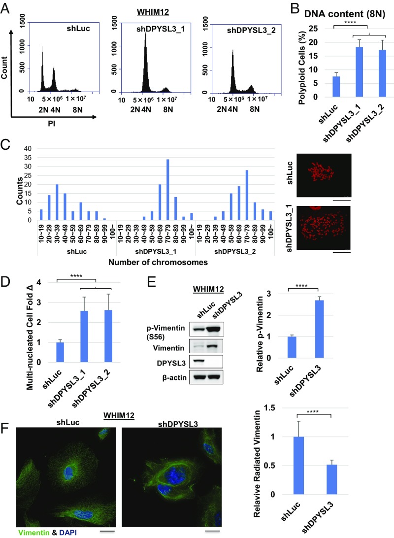
Fig. 3.
Multiple nucleation and polyploidy are induced in the absence of DPYSL3 in CLOW breast cancer. (A) Representative histograms depicting cell cycle distribution of stable WHIM12 shLuc, shDPYSL3_1, and shDPYSL3_2 cell lines by flow cytometry. (B) Quantification of 8N polyploid cells from A. WHIM12 shDPYSL3_1 and shDPYSL3_2 cells have a significantly higher percentage of 8N for polyploid cells compared with WHIM12 shLuc cells. ****P < 0.0001 (Student t test). (C) Mitotic spread of WHIM12 shLuc, WHIM12 shDPYSL3_1, and shDPYSL3_2 cells. Cells were stained with propidium iodide (PI), and the number of chromosomes was counted by using a confocal microscope or an Eclipse Ti microscope. (D) Bar graphs depicting fold change of multinucleated cells in WHIM12 shDPYSL3_1 and shDPYSL3_2 cells compared with shLuc, quantified from immunofluorescence. Data are averages from three independent experiments ± SEM. ****P < 0.0001 (derived from Student t test). (E, Left) Western blotting was performed with the indicated antibodies in WHIM12 shLuc and shDPYSL3 cells. (E, Right) Bar graphs depicting average phosphorylated vimentin levels from three independent experiments ± SEM. ****P < 0.0001 (derived from Student t test). (F, Left) Representative confocal microscopy immunofluorescence images of stable WHIM12 shLuc and shDPYSL3 cells stained with vimentin (pseudocolored in green) and DAPI (pseudocolored in blue). (F, Right) Bar graphs depicting average number of cells with radiated vimentin structure ± SEM from three independent experiments. ****P < 0.0001 (derived from Student t test).

