Fig. 4.
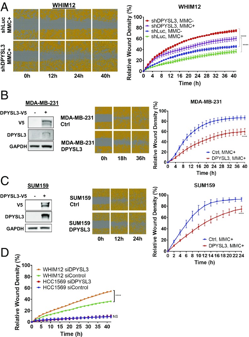
DPYSL3 inhibits migration in CLOW breast cancer. (A, Left) Representative cell migration images of stable WHIM12 shLuc and shDPYSL3 cells in the presence of mitomycin C (MMC+) at indicated times postwounding. Cells are pseudocolored orange to aid visualization. (A, Right) Quantification of relative wound density of indicated stable WHIM12 cell lines during the course of the wound recovery assay in the absence (MMC–) or presence of MMC. P value was determined by ANOVA. ****P < 0.0001 (Tukey’s multiple comparisons test). (B and C, Left) Western blots showing V5-tagged DPYSL3 expression in transfected MDA-MB-231 cells (B) and SUM159 cells (C). GAPDH was used as a loading control. (B and C, Center) Representative cell migration images of transfected cells from B and C, Left postwounding at the indicated time points. (B and C, Right) Relative wound density of indicated cell lines during the course of the wound recovery assay in the presence of MMC (MMC+). ****P < 0.001 (derived from Student t test comparing last time point measured). (D) Quantification from scratch wound healing assay of WHIM12 and HCC1569 transfected with siControl or siDPYSL3 in the presence of mitomycin C proliferation blocking agent. Data are averages from three independent experiments ± SEM. ****P < 0.001 (Bonferroni’s multiple comparisons test).

