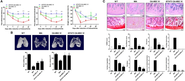
Figure 3.
Reduced severity by IV administration of iSTAT3 OA-MSCs in MIA-induced OA rats. (A) OA was induced in Wistar rats by IA injection of MIA. OA rats were injected IV with OA-MSCs (6 × 10⁵) or iSTAT3 OA-MSC (6 × 10⁵). OA-MSC were isolated of fat tissues obtain from each 3 person with OA. Pain behavior was assessed as mechanical hyperalgesia measured with a dynamic plantar esthesiometer and incapacitance meter, and was quantified as PWL and PWT (n = 3 per group). (B) Representative micro-CT images of the femoral condyles at 4 weeks in all groups. Object volume (Obj.V) and bone surface (%) measured in micro-CT images from femurs. (C) Histochemical analysis of femoral condyle and severity scores after IV administration of iSTAT3-OA MSCs in MIA-induced OA rats. Knee joint tissue samples were acquired at 4 weeks from wild-type (WT) rats, rats with MIA-induced OA, rats that received IV OA-MSCs, and rats that received IV iSTAT3 OA-MSCs, and were stained with H&E and Safranin O to evaluate the severity of inflammation and cartilage damage (*P < 0.05; **P < 0.01; ***P < 0.001).

