. 2018 Dec 17;7:e43224. doi: 10.7554/eLife.43224

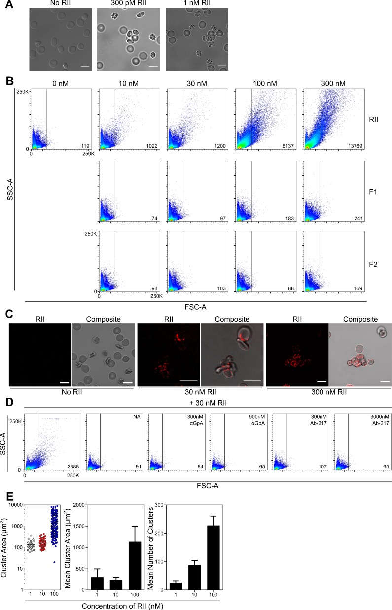
Figure 2. EBA-175 RII binds and promotes RBCs to form clusters through interactions with GpA.

(A) Clustered RBCs adhere to one another via RII at 300pM and 1 nM concentrations. Scale bars are 10 µm. (B) RII, but not the individual DBL domains F1 or F2, induces RBC clustering in a concentration dependent manner as observed by FSC-A. Frequency of events (clusters) out of 100,000 counts are located in bottom right corner of dot plot for each sample when gated according to the no protein control (top left). One representative biological replicate out of three is presented. (C) Clustered RBCs adhere to one another via RII at the interface between each red cell. Scale bars are 10 µm. (D) Inhibition of particle size shift of RBCs incubated with RII by neuraminidase (NA, panel 2), anti-GpA Ab (panel 3 and 4), and anti-RII Ab-217 as observed by FSC-A (panel 5 and 6). Frequency of events (clusters) out of 100,000 counts are located in bottom right corner of dot plot for each sample when gated according to the no protein control (top left). One representative biological replicate out of three is presented. (E) Analysis of cluster size for varying concentrations of EBA-175 RII under flow conditions of 1 dyn/cm2. Left hand panel shows representative data from one experiment. Each circle represents a single cluster area (µm2). Middle panel shows mean cluster size for each concentration, and right hand panel shows mean number of clusters per concentration for three independent experiments ± S.D.

Figure 2.

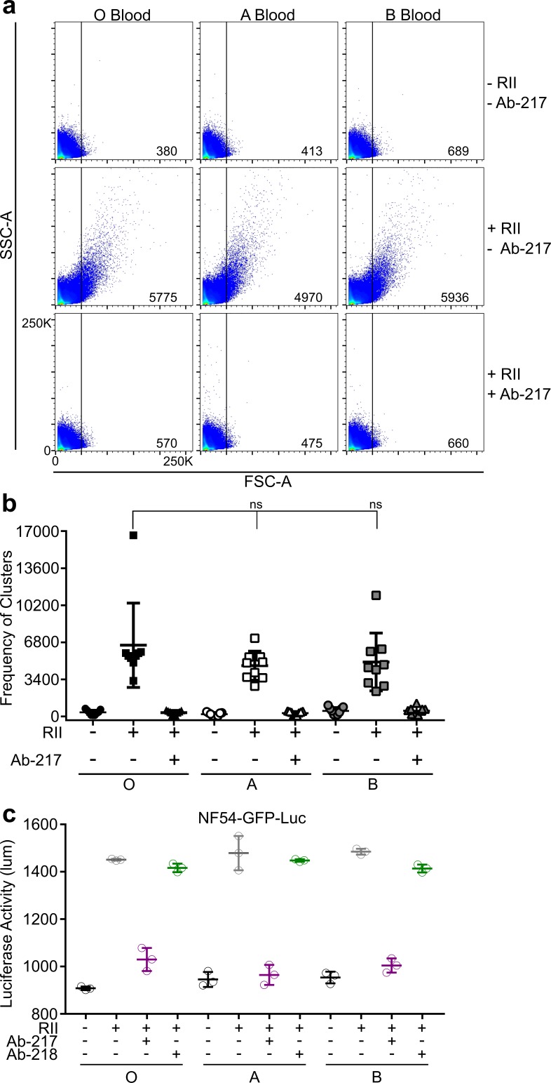
Figure 2—figure supplement 1. EBA-175-RII mediates similar levels of parasite growth in O, A, and B blood types.

Figure 2—figure supplement 1.

(A) RII induced RBC clusters in O, A, and B blood as observed by increasing FSC-A and SSC-A in the presence or absence of Ab-217. (B) The number of clusters was quantified by gating according to the control with no protein added, values are for nine biological replicates of one million counts each for each condition. Results are shown as ±SEM. ○=untreated RBCs, □=30 nM RII, Δ = 30 nM RII/300 nM Ab-217, black = O blood, white = A blood, dark gray = B blood. (C) RII-induced growth enhancement of NF54-GFP-luc parasites in O, A, and B blood types, in the presence Ab-217 or Ab-218, as assessed by luciferase assay. Data shown are mean ± SD of three technical replicates.
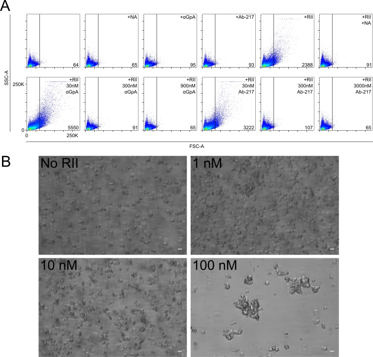
Figure 2—figure supplement 2. RII binds RBCs and forms stable clusters under flow conditions.

Figure 2—figure supplement 2.

(A) Inhibition of RII induced size shift of RBCs incubated with RII by neuraminidase, anti-glycophorin A Ab, and anti-RII Ab-217 and treatment controls without RII-175. FSC-A is correlated with the size or volume of an object while SSC-A is a measure of the internal composition of the object. Frequency of events (clusters) out of 100,000 counts are located in bottom right corner of dot plot for each sample when gated according to the no protein control (B) Representative images of RBCs under physiological flow conditions at various concentrations of RII. Scale bars are 10 µm.