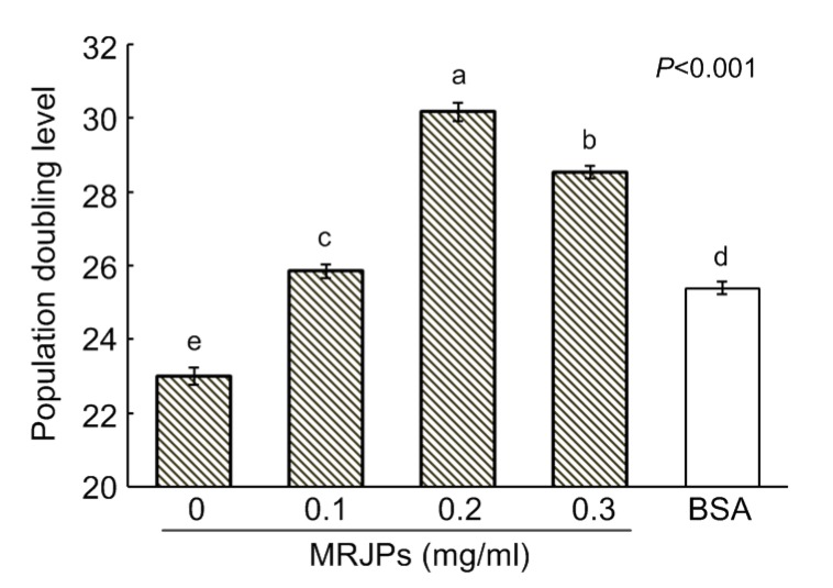
Fig. 2.
Lifespans of HFL-I cells supplied with different concentrations of MRJPs or BSA
The PDL represents the lifespan of HFL-I. Data are expressed as mean±SD (n=3). Different lowercase letters (a–e) above bars indicate a significant difference from each other at P<0.05 by Duncan’s multiple range tests

