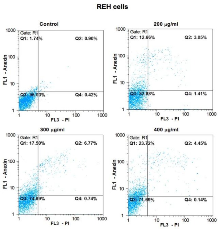
Figure 3.
Results from the induction of apoptosis by the B. aspera extract on REH cell line. The REH cells were exposed to intended concentrations of the B. aspera extract and then the induction of apoptosis after 48 h incubation was evaluated. Flow cytometry images, Q1, Q2, Q3 and Q4 represent early apoptosis, late apoptosis, live cells, and necrotic cells, respectively.

