
FIGURE 3.
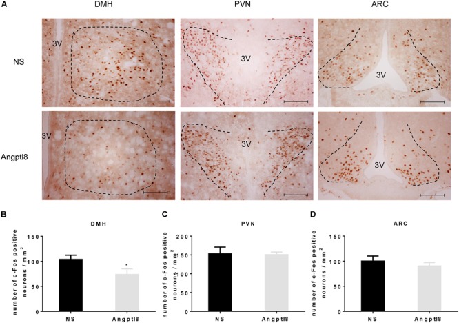
Peripheral Angptl8 decreased c-Fos-positive neurons expression in the DMH of the hypothalamus. (A) c-Fos-positive neurons change in hypothalamus 2 h after tail intravenous injection with NS and Angptl8. (B–D) The numbers of c-Fos-immunopositive cells in the DMH (n = 8), PVN (n = 6), and ARC (n = 8) are expressed as means ± SEM. ∗P < 0.05 relative to the NS control group. Scale bar in A = 100 μm.