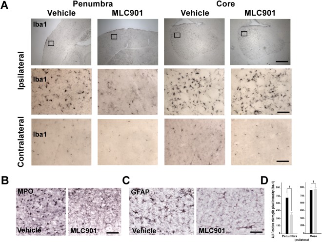
Figure 4.
MLC901 decreased microglial activation and neutrophil recruitment in the reperfused brain 24 hours following MCAO. (A–D) Representative immunohistochemical staining for Iba1 microglia marker (A) myeloperoxidase (MPO) (B) and GFAP astrocytic marker (C) in brains of vehicle- and MLC901-treated mice 24 hours after MCAO. (A) Representative views of Iba1 immunopositive cells in ischemic penumbra, ischemic core and contralateral control side of vehicle- and MLC901-treated mice. Diagram boxes indicate the regions where the images in middle and bottom panels at higher magnification were acquired (B,C) Representative immunostaining of MPO (B) and GFAP (C) in ischemic penumbra of vehicle- and MLC901-treated mice. (D) Quantitation of Iba1 expression (AU positive microglia pixel intensity) in ischemic penumbra and ischemic core of vehicle- and MLC901-treated mice. Scale bar, 20 μm (top panels), 50 μm (middle and bottom panels) (n = 6 per experimental group).

