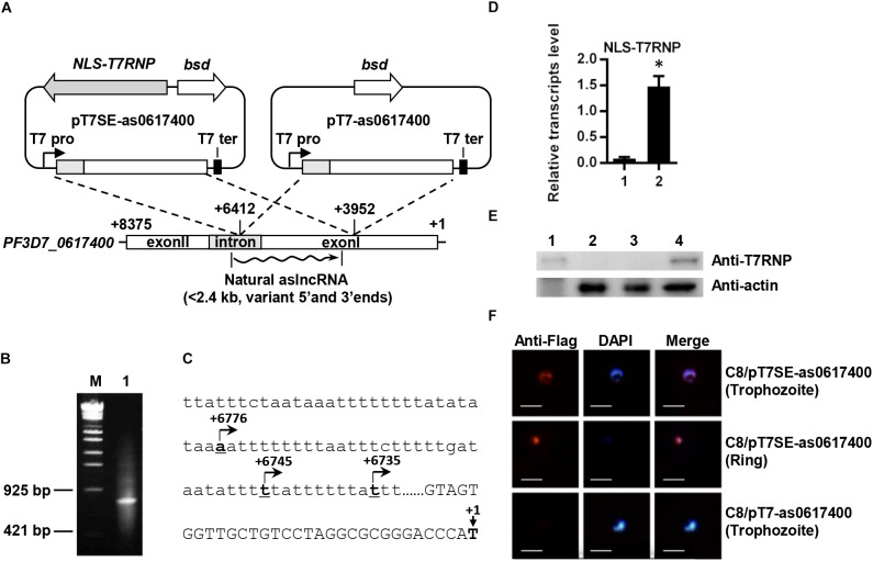
FIGURE 1.
Schematic diagram of plasmids for expressing the var aslncRNA and 5′ end identification of PF3D7_0617400 aslncRNA. (A) Schematic diagram of pT7SE-as0617400 and pT7-as0617400. The translation initial site of ATG of PF3D7_0617400 is marked as +1 and the stop codon is marked with +8375, the ∼2.5 kb var fragment (from +6412 to +3952) was amplified and inserted into pT7SE and pT7, respectively. The wavy line is the natural var aslncRNA. T7 pro, T7 promoter; T7 ter, T7 terminator. (B) 5′ end identification of PF3D7_0617400 aslncRNA. PCR products of 5′ end RACE obtained according to the standard manuals. M: the DNA marker λ-EcoT14 I digest (TAKARA), lane1: PCR products of 5′ RACE. (C) The PF3D7_0617400 aslncRNA transcriptional start sites. The translation start site of PF3D7_0617400 is marked as +1 on the schematic diagram. Capital letters are the coding sequence of the var exonI and the lower letters are intron sequence. The transcriptional start sites of the aslncRNAs are marked with underlined numbers and arrows. The underlined letters locations are displayed relative to translation start site, and the arrows indicate the aslncRNA transcription directions. (D) RT-qPCR quantification of the NLS-T7RNP transcription in C8/pT7- as0617400 (1) and C8/pT7SE-as0617400 (2). Relative transcripts numbers are normalized to serine-tRNA ligase gene (PF3D7_0717700). ∗P < 0.05 by paired two-tailed Student’s t-test. The RT-qPCR results are representative of three independent experiments with data indicating the mean +SD. (E) Western blot analysis of NLS-T7RNP expression in C8/pT7-as0617400 and C8/pT7SE-as0617400. The NLS-T7RNP is detected by anti-T7RNP antibody. Lane1: recombinant NLS-T7RNP expressed in E. coli BL21, Lane2: the wild type parasite 3D7 strain C8, Lane3: C8/pT7-as0617400, Lane4: C8/pT7SE-as0617400. (F) The subcellular localization of NLS-T7RNP in the transformant C8/pT7SE-as0617400 and C8/pT7-as0617400. NLS-T7RNP is marked with anti-Flag antibody (red) and is the nuclear marked with DAPI (blue). The scale bar is 25 μm.

