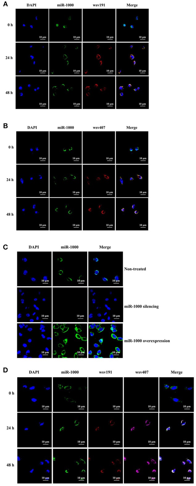
Figure 6.

Co-localization of miR-1000 and its target mRNAs in shrimp in vivo. (A) Co-localization of miR-1000 and wsv191 mRNA in shrimp hemocytes. Shrimp were infected with WSSV. At different time points post-infection, miR-1000, wsv191 mRNA, and nuclei of hemocytes were respectively detected with FAM-labeled miR-1000 probe (green), Cy3-labeled wsv191 probe (red), and DAPI (blue). Scale bar, 10 μm. (B) Co-localization of miR-1000 and wsv407 mRNA in shrimp hemocytes. Shrimp hemocytes were examined by FAM-labeled miR-1000 probe (green) and Cy5-labeled wsv407 probe (red). Scale bar, 10 μm. (C) Specificity of miR-1000 probe. Shrimp were injected with AMO-miR-1000 or miR-1000 mimic. At 48 h after injection, miR-1000 was detected with FAM-labeled miR-1000 probe (green). Scale bar, 10 μm. (D) Evaluation of co-localization of miR-1000, wsv191 mRNA, and wsv407 mRNA in shrimp hemocytes. The WSSV-infected shrimp hemocytes were subjected to fluorescence in situ hybridization with FAM-labeled miR-1000 probe (green), Cy3-labeled wsv191 probe (red), and Cy5-labeled wsv407 probe (pink), respectively. Scale bar, 10 μm.
