Figure 1.
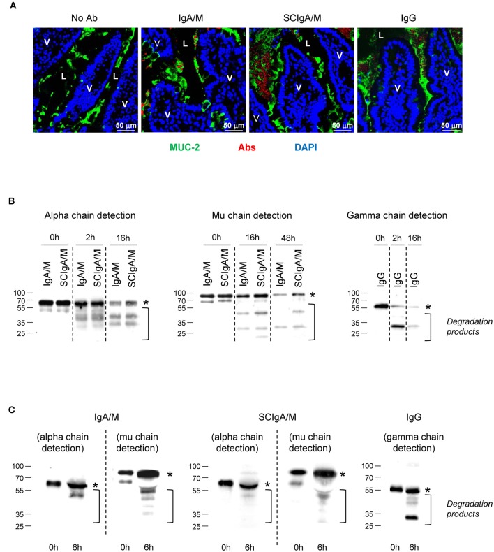
Location and stability of Ab preparations in the gastrointestinal tract. (A) Tissue sections prepared from mouse ligated intestinal loops 1.5 h post-administration with either Cy5-labeled IgA/M, SCIgA/M, or IgG appearing in red. Sections were further stained with anti-MUC-2 antiserum and DAPI to detect mucus and cell nuclei prior to analysis by laser scanning confocal microscopy. Examples of typical results of more than five samples are shown. L, Lumen. Magnification, x 40 for all panels. (B) In vitro digestion patterns observed at three time points of IgA/M, SCIgA/M, and IgG incubated with intestinal washes, as assayed by immunodetection of the respective heavy chain under reducing conditions. In (B,C), the position of the intact alpha, mu, and gamma chains is indicated by an asterisk. (C) IgA/M, SCIgA/M, or IgG incubated for 6 h in a ligated intestinal loop were recovered as described in Materials and Methods, and analyzed as under (B).

