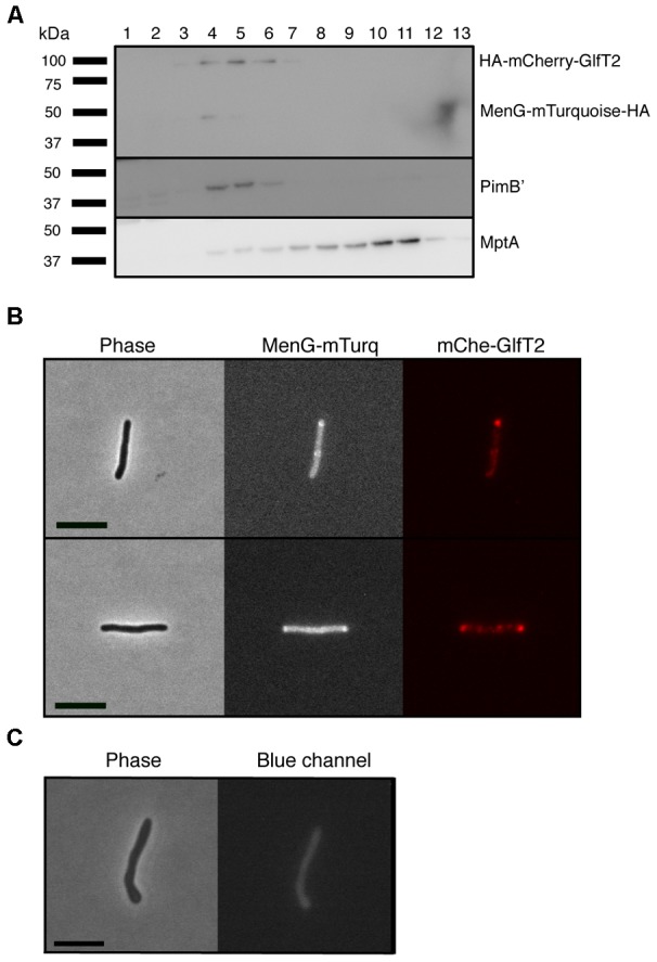
FIGURE 3.

Co-localization of MenG with the IMD-associated protein GlfT2. (A) Sucrose density fractionation of strain expressing HA-mCherry-GlfT2 (100 kDa) and MenG-mTurquoise-HA (50 kDa). The epitope-tagged proteins were detected by anti-HA antibody. PimB’ (41 kDa) and MptA (54 kDa), respectively, indicate the IMD and PM-CW fractions. (B) Fluorescence microscopy showing localization of both MenG-mTurquoise-HA and HA-mCherry-GlfT2 at the pole of growing M. smegmatis cells. (C) Autofluorescence of wild-type M. smegmatis on blue channel observed under the identical image acquisition setting as in panel B. Scale bar = 5 μm. All experiments were done more than twice and representative data are shown.
