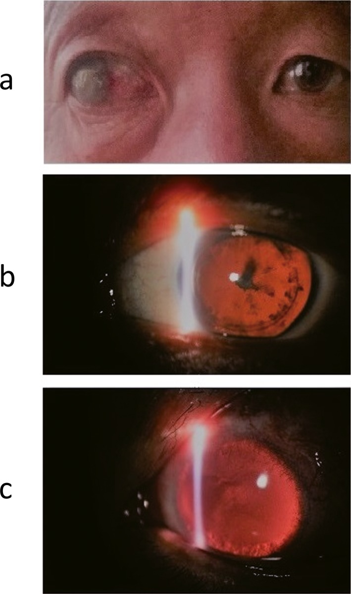
Figure 2.

Eye pictures of partial members of the Family 1 (provided by the proband). Part a is the binocular photograph of the proband's father (II2, Figure 1) as control, whose right eye is traumatic blindness and left eye is normal; Part b is the slit‐lamp biomicroscopy photography of the left eye of the patient (III5, Figure 1) as proband, who combined with cataract before cataract surgery; Part c is the slit‐lamp biomicroscopy photography of the left eye of the patient (IV1, Figure 1)
