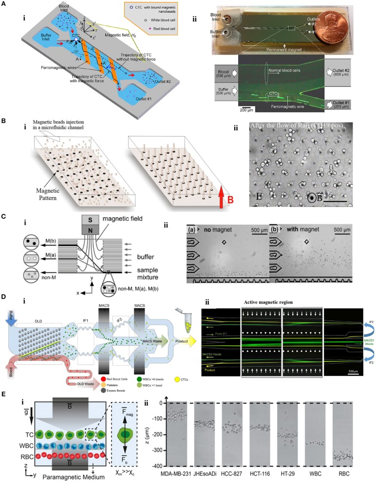
Figure 3.
Microfluidic tumor cell separation devices. (A) Separation of magnetically-labeled target cells. (i) Design and (ii) optical micrograph of the positive tumor cell (SKBR-3) enrichment device using anti-EpCAM-coupled magnetic nanoparticles. Reprinted with permission from Kim et al. (2013b). Copyright (2013) American Chemical Society. (B) In situ magnetic labeling of tumor cells (Ephesia). (i) The magnetic beads are located on ferrofluid dots in the microfluidic channel to create a self-assembled magnetic bead array under the applied magnetic field. (ii) Cells are captured on the magnetic bead array during the sample flow. Reprinted from Saliba et al. (2010). (C) Separation of tumor cells (HeLa) based on MNP uptake extent. (i) The schematic illustration of the device. (ii) HeLa cells are deflected from the laminar flow according to their magnetic load. Reprinted with permission from Pamme and Wilhelm (2006). Copyright (2006) The Royal Society of Chemistry. (D) Separation of cancer cells by depleting other cells. (i) A monolithic microfluidic chip for negative enrichment of tumor cells through the depletion of magnetically-labeled WBCs. In the deterministic lateral displacement (DLD) part, RBCs, platelets, and free beads are eliminated. Following the inertial focusing 1 (IF1), magnetophoresis (MACS1) is applied to deplete magnetic labeled WBCs having more than ~6 beads. Another set of inertial focusing (IF2) and magnetophoresis (MACS2) is applied to deplete WBCs that contain at least one magnetic bead. (ii) Image of cell streaks captured using fluorescence microscopy on different areas of the chip. Green and yellow colors represent WBCs and CTCs, respectively. Reprinted from Fachin et al. (2017). (E) Tumor cell separation using cells' intrinsic properties in a paramagnetic solution. (i) The design of the label-free magnetophoresis platform (MagLev). (ii) Alignment of tumor cells at different heights (z-axis) in the device. Reprinted from Durmus et al. (2015).

