Figure 1.
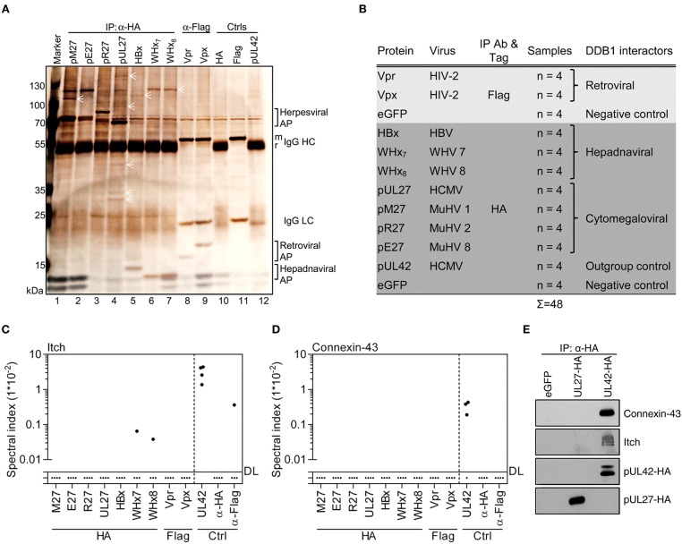
Viral DDB1-interacting proteins exhibit distinct co-precipitation profiles. (A) Accessory proteins implicated in the exploitation of DDB1 were expressed in HEK293T cells. Lysates were generated and subjected to IP analysis using the indicated antibodies. Retrieved proteins were visualized by silver staining of SDS gels. Symbols highlight proteins representing partially overlapping and specific interactions. Control IPs were performed using cells transfected with pUL42- and eGFP-expressing plasmids. (B) Overview of the experimental conditions for IP coupled to mass spectrometry (MS). The indicated proteins were analyzed by IP as described in (A) but instead of using silver stained gels, (co-)precipitated proteins were identified and quantified by MS in four biological replicates. Spectral indexes were calculated for Itch (C) and Connexin-43 (D). (E) The indicated proteins were expressed in HEK293T cells. Lysates were subjected to IP using an HA-specific antibody and immunoblot analysis was performed for the indicated proteins.

