
Fig. 2.
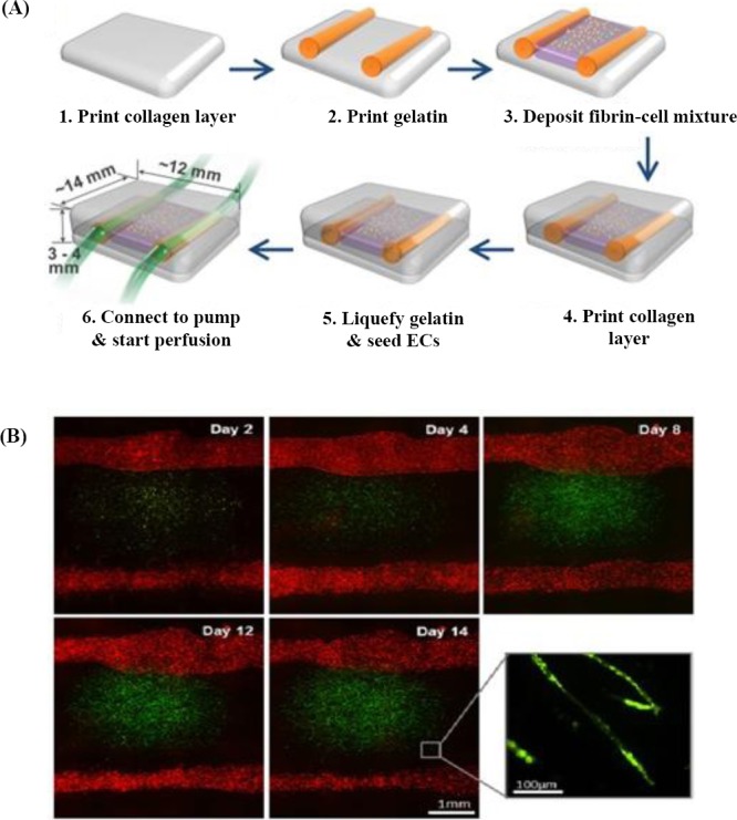
Extrusion printing method. (A) A schematic picture of two large channel structures and deposition of fibrin and cell, which were developed using the 3D bioprinter. (B) Florescent microscopic images of two large channels, and the mixture of fibrin cell placed between channels. Green dots show green fluorescent protein-ECs that were cultured within fibrin, and red dots indicate red fluorescent protein-ECs that were seeded on the two large channels[55].