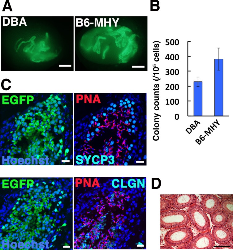
Fig. 2.
Functional analysis of MHY-GS cells by spermatogonial transplantation. (A) Macroscopic appearance of a recipient testis transplanted with MHY-GS cells. Green fluorescence indicates colonies originating from transplanted GS cells. (B) Colony counts (n = 19–21). (C) Immunostaining of recipient testis for SYCP3, CLGN, and PNA. (D) Histological appearance of epididymis of leuprolide-treated mouse. Bar = 1 mm (A), 20 μm (C), 100 μm (D). Counterstain: Hoechst 33342 (C), H & E (D).

