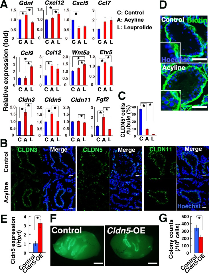
Fig. 4.
Analysis of acyline-treated mouse testes. (A, B) Real-time PCR (A; n = 6) and immunostaining analyses (B) of acyline- and leuprolide-treated testis. Recipient mice were treated with busulfan to remove endogenous spermatogenesis 1 month before analysis. (C) Quantification of tubules with CLDN5+ Sertoli cells. Cells in 10 tubules were counted. (D) Functional assessment of the BTB in acyline-treated mouse testis. Recipient mice were injected interstitially with biotin (green). (E) Real-time PCR analysis of busulfan-treated testes that received lentivirus that overexpress Cldn5 (n = 3). Samples were recovered 2 days after microinjection. (F) Macroscopic appearance of recipient testis that received GS cell transplantation. (G) Colony counts (n = 18 for Cldn5; n = 16 for control). Bar = 20 μm (B), 50 μm (D), 1 mm (F) . Counterstain: Hoechst 33342 (B, D). Asterisk indicates statistical significance (P < 0.05).

