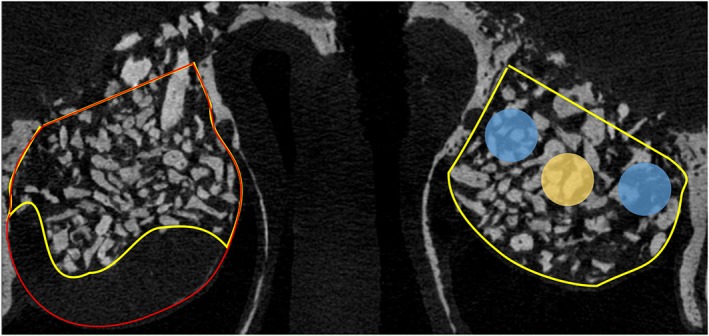
Figure 1.

Drawing exemplifying the volumes and regions analyzed. The volume included in the analyses of the augmented sinus is outlined in yellow. The volume occupied by the inner collagen membrane was excluded from the analyses (outlined in red). Three interpolated cylindrical regions of 1 mm in diameter were used, located close to the medial and lateral bony walls (Walls region; light blue circle) and in the middle of the augmented volume (Middle region; yellow circle)
