FIGURE 1.

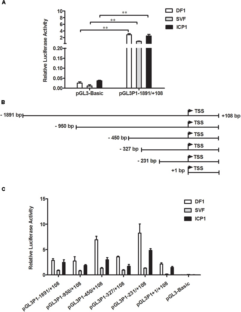
Characterization of the chicken PPARγ gene P1 promoter. (A) Luciferase activity of the P1 promoter reporter construct pGL3P1-1891/+108 in DF1, SVF, and ICP1 cells. Cells were transfected with pGL3P1-1891/+108 along with the pRL-TK Renilla luciferase vector by using Lipofectamine 2000 reagent, and luciferase activity was determined at 48 h after transfection. (B) Schematic diagram of the reporter constructs of the P1 promoter and its 5′ truncation mutants. The transcription start site (TSS) of chicken PPARγ1 is represented by the bent arrow. The positions are numbered relative to the TSS. (C) Truncation analysis of the P1 promoter in DF1, SVF, and ICP1 cells. The indicated P1 promoter constructs and pRL-TK vector were cotransfected into DF1, SVF, and ICP1 cells. Luciferase activity was detected 48 h after cotransfection. The pRL-TK vector was used for normalization of transfection efficiency. All data represent the mean ± SE. Statistical significance was determined by Student’s t-test. ∗∗p < 0.01.
