Fig. 6.
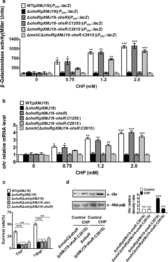
Positive regulation of ohr by OhsR. a β-Galactosidase analyses of ohr promoter activities by using the transcriptional Pohr::lacZ chromosomal fusion reporter expressed in indicated strains exposed to stress conditions for 30 min.***P < 0.001; **P < 0.01; *P ≤ 0.05. b Quantitative RT-PCR analyses of ohr expression in indicated strains exposed to stress conditions for 30 min. Results were the average of four independent experiments; the standard deviation was indicated by bars. ***P < 0.001; **P < 0.01; *P ≤ 0.05. c Survival rates for five relevant strains after challenged with 11 mM CHP and 20 mM t-BHP. Mean values with standard deviations (error bars) from at least three repeats were shown. ***P ≤ 0.001; **P < 0.01. d The protein levels of Ohr in ΔohsR(pXMJ19-ohsR:C261S) and ΔmshCΔohsR(pXMJ19-ohsR:C261S), strains with or without CHP treatment. Lysates from stationary phase bacteria exposed to 1.5 mM CHP for 1 h were resolved by SDS-PAGE, and Ohr was detected by immunoblotting using specific anti-Ohr antibody. For the pellet fraction, RNA polβ was used as a loading control. Similar results were obtained in three independent experiments, and data shown are from one representative experiment done in triplicate (Left panel). Relative quantified data for protein levels by Image Lab (Right panel). Quantified protein expression of western blots in d. Densities of proteins were all justified with α-RNA polβ (beta chain of RNA polymerase). Relative density ratios of Ohr in the ΔohsR(pXMJ19-ohsR:C261S) strain without stress were set at a value of 1.0. Data shown were the averages of three independent experiments, and error bars indicate the SDs from three independent experiments. **P < 0.01; *P < 0.05. n.s. not significant

