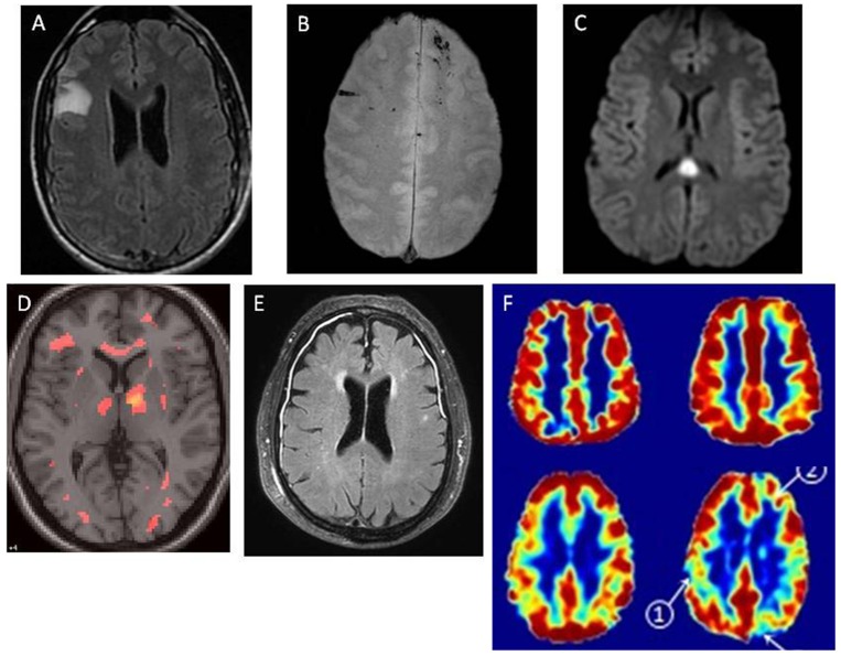
Figure 3.
Magnetic resonance images of patients with post-concussion symptoms. MRI findings in patients with mTBI, demonstrating multiple pathologies. In each case, cranial CT was normal. MRI was obtained within 48 h on injury. (A) Right frontal non-hemorrhagic contusion, noted on FLAIR image. (B) Linear microhemorrhages in left and right frontal lobes, noted on T2* image. (C) Diffuse axonal injury lesion in splenium of corpus callosum, with restricted diffusion noted on DWI image. (D) Diffuse axonal injury, with multifocal lesions noted on diffusion tensor imaging (DTI). (E) Traumatic meningeal enhancement of subdural effusions, noted on post-gadolinium FLAIR image. (F) Traumatic microvascular injury.
- - Top row represents a single healthy control. Bottom row represents a single TBI patient.
- - Left column: Cerebral Blood Flow (CBF), assessed by arterial spin labeling.
- - Right column: Cerebrovascular reactivity (CVR) assessed using BOLD response to hypercapnia.
Credit for figures: Figures A, B, C, E: Larry Latour, PhD, NINDS/NIH; D: Carlos Marquez de la Plata, PhD, University of Texas at Dallas; F: Franck Amyot, PhD, Uniformed Services University of the Health Sciences.

