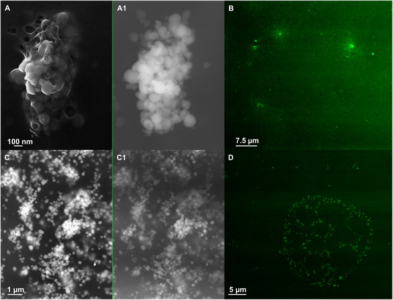
FIGURE 6.
Scanning electron microscopy (SEM) of SeNPsSeITE02-G_e [A (In Lens detector), A1 (detector for back scattered electron)] and SeNPsSeITE02-P_e [C (In Lens detector), C1 (detector for back scattered electron)]. Confocal laser scanning microscopy (CLSM) of SeNPsSeITE02-G_e (B) and SeNPsSeITE02-P_e (D) labeled with the lipophilic tracer DiOC18(3).

