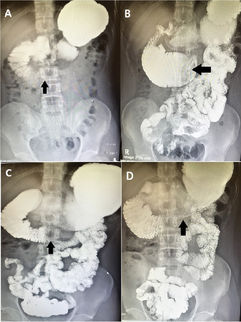
Figure 1.

(A-D) Upper gastrointestinal series with small bowel follow-through. Oral contrast was administered. Note the distended stomach and proximal duodenum as well as the narrow distal duodenum. There is abnormal dilation of the third part of the duodenum, measuring 7.1 cm in diameter, as well as delayed passage of contrast into the distal small bowel: features suggestive of a partial distal duodenal obstruction.
