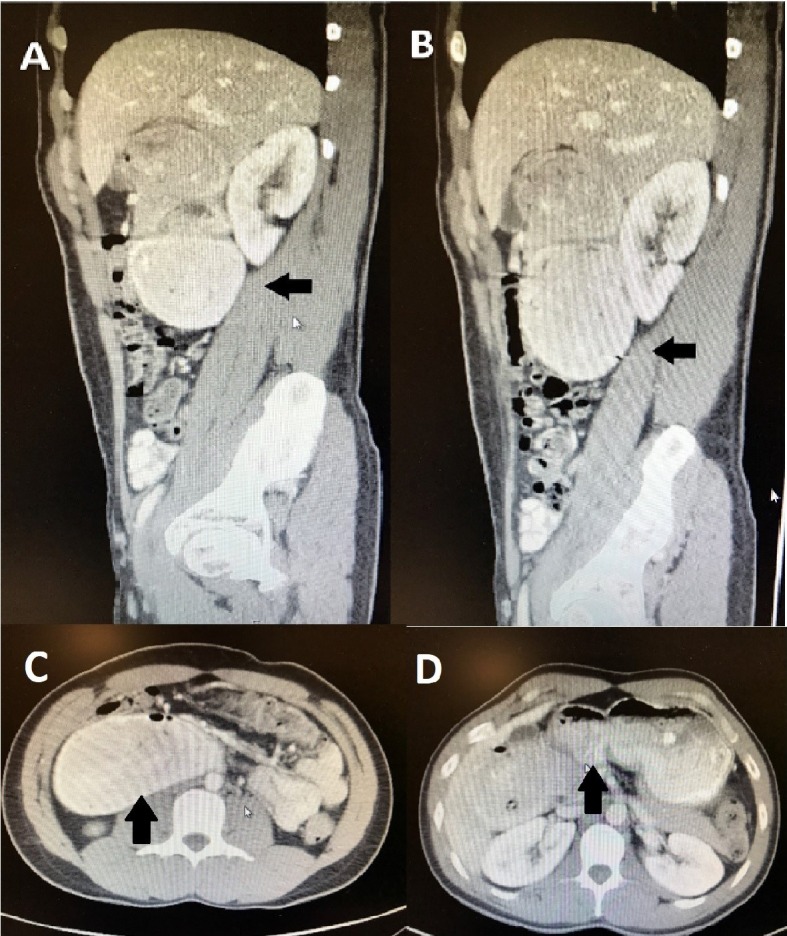
Figure 2.

(A-D) Abdominal CT scan. Oral contrast was administered. The patient was discovered to have marked dilation of the second and third parts of the duodenum as well as marked narrowing of the distal third and fourth parts, which was consistent with high-grade partial small bowel obstruction at the distal duodenum.
