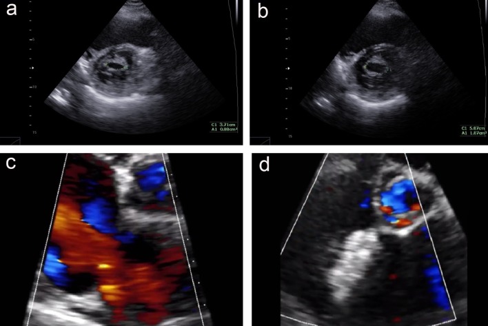
Figure 3.
The echocardiography exam before and 12 months after procedure. (a) Mitral valve at parasternal short axis view, before mitral valvuloplasty; (b) mitral valve at parasternal short axis view, after mitral valvuloplasty; (c) the color Doppler echocardiography at parasternal short axis view shows an ASD with left to right shunt; (d) the Cocoon Septal Occluder is in good position without residual shunt.

