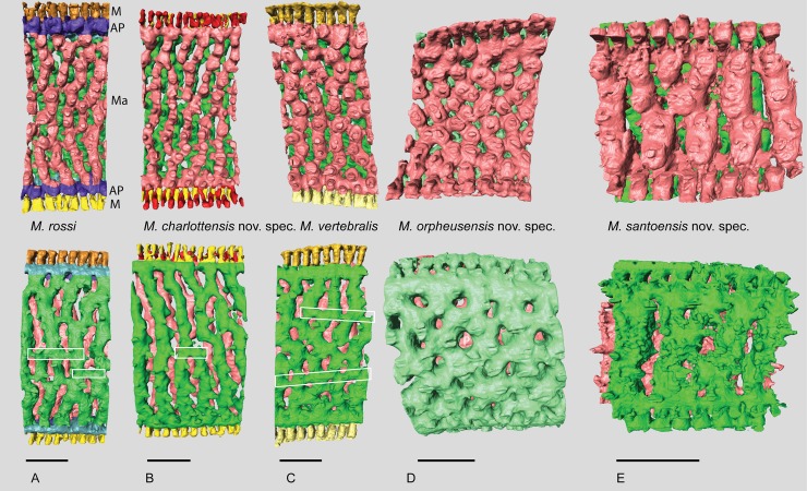
Fig 5. Micro-computed tomography of Marginopora with the shell removed, showing the interseptal space of two subsequent chambers at a thickness of ~10 stolon planes, demonstrating the structural differences between the five species.
The top row is projected looking from the inside out, the bottom row shows the same chambers looking from the outside in. Pink chamber is the innermost, green chamber the outermost of the reconstructed chambers. In A-C the marginal chambers have been highlighted in yellow. A) M. rossi, showing frequent annular connections (white squares) of 2–3 adjacent chamberlets. Note the three way differentiation within the chamber: marginal chamberlets (M, yellow) connection to the annular passage of the current and preceding whorl on either side of the shell, the annular passage (AP, blue and purple) which is partitioned by septula, and the marginoporid structure (Ma) containing the median chamberlets and the radially oblique stolon planes (RGM.1352065). B) M. charlottensis nov. spec. showing regular chamberlet width and the very rare annular connections (white squares), resulting in a very regular looking shell (G466207). C) M. vertebralis showing the irregular shape of the chamberlets, the frequent annular connections sometimes connecting up to 5 adjacent chamberlets (white squares) (RGM.1352105). D) M. orpheusensis nov. spec. showing the high interconnectedness of the chamberlets. Apart from the radially oblique stolon planes, chamberlets are also connected by stolon planes within the chamberlets at ~45° to the annular passage (G466208). E) M. santoensis nov. spec. showing the regular chamberlet shape and the low level of interconnectedness (RGM.1352067). Scale bars are approximately 200 μm.

