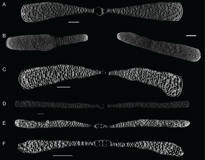
Fig 6. Virtual vertical thin sections through the A-forms of Marginopora studied here.
A) M. rossi, small specimen, showing the embryonic apparatus and the gradual increase in thickness (RGM.1352063; scale bar 500 μm). B) M. rossi, large specimen, which lost the embryonic apparatus and initial whorls (RGM.1352066; scale_bar 1 mm). C) M. vertebralis (RGM.1352103; scale bar 500 μm). D) M.orpheusensis nov. spec. (RGM.1352059; scale bar 500 μm). E) M. charlottensis nov. spec. (RGM.1352045); Scale bar 500 μm). F) M. santoensis nov. spec. (RGM.1352101; Scale bar 500 μm).

