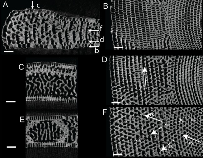
Fig 9. Virtual thin sections of Marginopora vertebralis A-form from Newton Island (specimen RGM.1352105).
A) Radial virtual vertical thin sections showing the positions of B-F. B) Virtual horizontal section through the lateral chamberlets. C) Virtual vertical thin section perpendicular to A, in the outer part of the shell, showing the bifurcating and irregular interseptal ridges. D) Virtual horizontal section through the annular passage (ap). E) Virtual vertical thin section perpendicular to A, in the inner part of the shell, showing few bifurcations and more regular interseptal ridges than in C. F) Virtual horizontal section through the marginoporid structure showing abundant annular connections (ac) and the alternating directions of the stolon passage (rs). Scale bars 100 μm.

