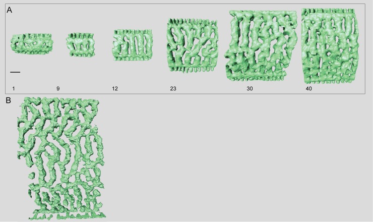
Fig 10. Micro-computed tomography of Marginopora rossi with the shell removed, showing the interseptal space.
A) chamber 1, 9, 12, 23, 30, and 40 of specimen RGM.1352065; and B) one of the thickest chambers of RGM.1352066. This specimen is missing its central part, so it is not possible to provide an exact chamber number. Note the fully developed chamber structure in chamber 1, as well as the long interseptal ridges even in the thickest chambers. Scale bar is approximately 100 μm).

