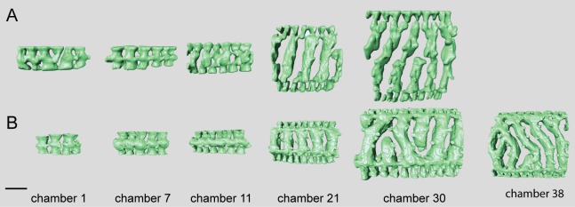
Fig 17. Micro-computed tomography of Marginopora santoensis nov. spec with the shell removed, showing the interseptal space.
A) the first, seventh, 11th, 21th, and 30th chamber of specimen RGM.1352068; B) the 7th, 11th, 21th, 30th and 38th chamber of specimens RGM.1352070. Note the increase in complexity within specimens, and the similarity in the order of changes between the specimens, but in the thicker specimen RGM.1352068) these changes occur in earlier chambers. Scale bar is approximately 100 μm.

