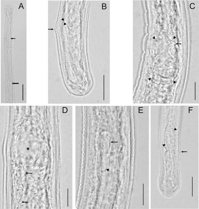
Fig 3. Imaging of O. volvulus larvae recovered from various models.
A) Anterior end of female larva showing overall shape, location of nerve ring (small arrow) and vulva (large arrow). [Scale bar = 50 μm] B) Female tail, lateral view, showing overall shape and rectum (arrow heads) and anal opening (arrow). [Scale bar = 15 μm] C) Developing ovejector (arrowheads), lateral view, showing attachment to body wall and beginning of vulva (arrow). [Scale bar = 10 μm] D) Developing ovejector, dorsal-ventral view, showing the relative large cavity (asterisk) and the developing tube (arrows). [Scale bar = 10 μm] E) Male larva at approximately mid-body showing the testis (arrow), which is C-shaped, curved posteriorly, and has begun to grow posteriorly (arrowhead). [Scale bar = 10 μm] F) Male tail, lateral view showing overall shape. One developing spicule pad (arrowheads) is clearly visible as is the anal opening (arrow). [Scale bar = 15 μm].

