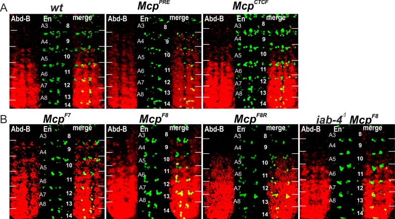
Fig 3. Expression of Abd-B in Mcp replacement embryos.
(A) Abd-B expression in wt, McpPRE and McpCTCF embryos. (B) Abd-B expression in McpF7, McpF8, McpF8R and iab-4Δ McpF8 embryos. Each panel shows an image of the embryonic CNS of stage 14 embryos stained with antibodies to Abd-B (red) and Engrailed (En, green). En is used to mark the anterior boundary of each parasegment. White horizontal bars approximately delimit parasegment/segment boundaries. Parasegments numbered from 9 to 14 on the right side of the panels; approximate positions of segments are shown on the left side of the wild type (wt) panel and marked A4 to A8. The wild type expression pattern of Abd-B in the embryonic CNS is characterized by a stepwise gradient of increasing protein level from PS10 to PS14. The McpF8 or McpF7 embryos have similar low Abd-B expression in PS9 and PS10. The Abd-B expression in PS9 is absent in iab-4Δ McpF8 and McpF8R embryos.

