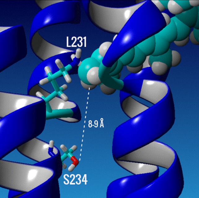
Fig 8. Close up of the retinal binding pocket in a homology model of GPR-DNFS.
The K231 and the mutant S234 on helix G (central blue helix in picture) are selectively displayed as stick models of the amino acid side chains, while the A1 retinal chromophore is displayed as a space filled residue in cyan. Dashed line shows estimated distance between the hydroxyl oxygen of S234 and the C15 carbon of the SB of the A1 retinylidene chromophore. The homology model was generated using sensory rhodopsin II (SRII) as a template (PDB 1H2S) [102] with the program YASARA (www.yasara.org) as described previously in [35] and chapter 2 of ref. [85].

