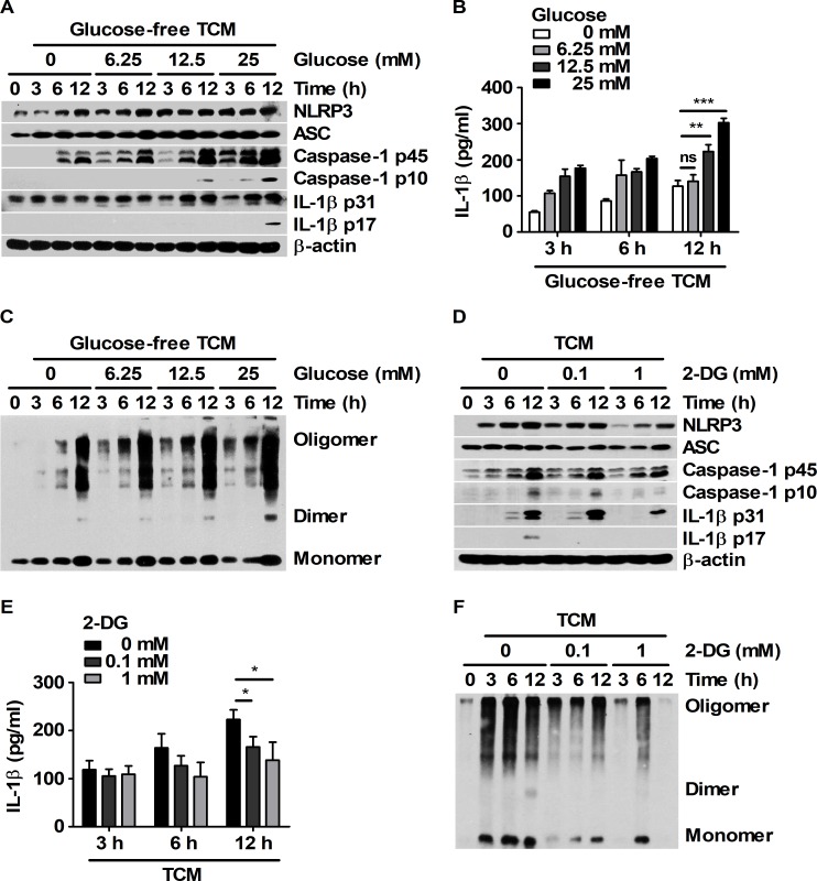
Fig 2. Mature IL-1β was generated by TCM stimulation through glucose-dependent inflammasome activation in BMDMs.
(A-C) BMDMs were cultured with 25% TCM mixed with glucose-free media supplemented with 0, 6.25, 12.5 or 25 mM glucose for 3, 6, and 12 h. (A) Western blots for NLRP3, ASC, caspase-1, and IL-1β were performed. β-actin was used as the control. (B) Secreted IL-1β levels in the supernatant were analyzed by ELISA. (C) ASC oligomerization was analyzed by DSS chemical crosslinking assay. (D-F) BMDMs were pre-treated with DMSO or 2-DG (0.1 or 1 mM) for 1 h and then treated with 25% TCM for 3, 6, and 12 h. (D) NLRP3, ASC, caspase-1, and IL-1β levels in the harvested cells were measured by western blot. β-actin was used as the control. (E) IL-1β levels were analyzed by ELISA. (F) ASC oligomerization was analyzed by DSS chemical crosslinking assay. The bars and error bars represent the mean ± SD; *, P < 0.05; **, P < 0.01; ***, P < 0.001; ns, not significant.

