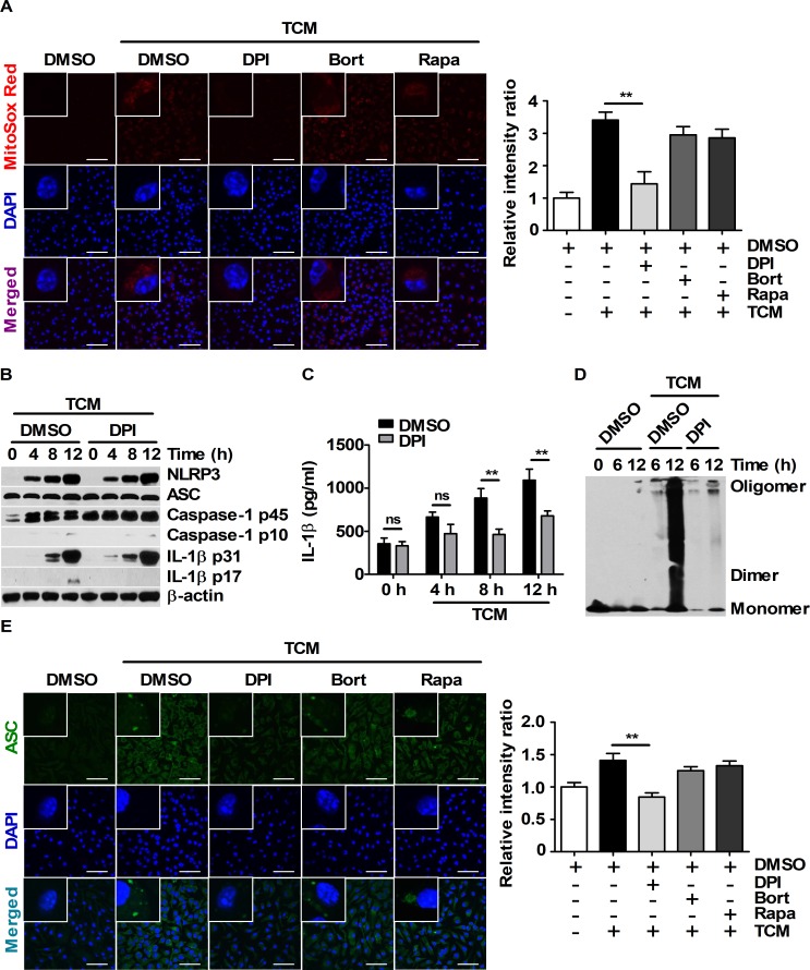
Fig 5. Enhanced ASC-ASC interactions are related to intracellular ROS levels in TCM-stimulated BMDMs.
(A and E) BMDMs were pre-treated with DMSO, 20 μM DPI, 5 μM bortezomib or 10 nM rapamycin for 1 h. Then, the cells were stimulated with 25% TCM. (A) Mitochondrial ROS localization in BMDMs stimulated with DMEM or 25% TCM for 6 h was visualized by MitoSOX Red staining. Nuclei (blue) were stained with DAPI. Scale bars, 50 μm. (B-D) BMDMs were pre-treated with DMSO or 20 μM DPI for 1 h and then stimulated with 25% TCM for the indicated times. (B) Protein levels of NLRP3, ASC, caspase-1, and IL-1β were measured by western blot. (C) The supernatant obtained in (B) were evaluated by ELISA. (D) ASC oligomerization was analyzed by DSS chemical crosslinking assay. (E) ASC specks in BMDMs stimulated with 25% TCM were visualized by immunostaining with an anti-ASC antibody (green). Nuclei were stained with DAPI (blue). Scale bars, 50 μm. The bars and error bars represent the mean ± SD; *, P < 0.05; **, P < 0.01; ***, P < 0.001; ns, not significant.

